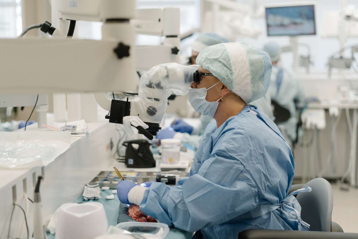
Through advanced equipment, instruments, and materials, conventional surgery is evolved into endodontic microsurgery. As a result, specialists can also produce predictable outcomes and favorable patient responses.
In
clinical terms, these procedures are referred to as treatments performed on an
infected tooth’s root apices. Oral surgeons, endodontists, and general
practitioners use various techniques to seal the root end with filling
materials.
Through
all these advances, patients get an enhanced solution to retain teeth that need
extraction otherwise.
Critical
differences
In
most cases, invasive endodontic intervention is not the first option for
treating an affected tooth. Specialists perceive it as tedious since they have
to locate anatomical structures, such as blood vessels, foramina, and necrotic
tissues. Thus, locating and cleaning damaged areas with traditional techniques
is challenging.
Endodontic
microsurgery involves the use of microscopic magnification and illumination to
achieve precision and predictability. Therefore, surgeons can precisely
identify root apices and resection angles without any troubles. Furthermore,
the use of ultrasonic instruments ensures an increased survival rate for
root-end preparations.
Use
of microscopes
One
of the most significant developments in endodontics is the use of operating
microscopes for surgical procedures. It provides better visual access to an
operating field for ensuring high treatment quality.
Notable
benefits
Endodontists
use microscopes to achieve a high magnification of small anatomical details.
For instance, they can identify and manage lateral canals with added precision.
Furthermore, it preserves the natural integrity of root ends.
Another
advantage is surgeons get to remove diseased tissues properly, even with
conventional protocols. As a result, they can eliminate any risks of
reinfections after a tooth filling.
Specialists
get to identify between the bone and root tips even with methylene blue
staining. Using microscopes allows them to intervene at higher magnification,
so faster healing is promoted.
Since
clinicians can inspect apices directly with these devices, they can reduce the
number of radiographs needed for endodontic treatment. This advantage further
helps to save time need for office trips.
Lastly, endodontists can use digital video recordings to educate patients. For endodontic microsurgery, patient preparation proves critical for successful outcomes.

Comments
Post a Comment